
The Discovery, Diagnosis, Pathophysiology & Treatment of Babesiosis
First question: Who discovered it?
Answer (just in case you, like me, didn’t know):
Victor Babeș (1854 - 1926) was a Romanian physician, bacteriologist, academician and professor. One of the founders of modern microbiology, Victor Babeș is the author of one of the first treatises of bacteriology in the world – Bacteria and their role in pathological anatomy and histology of infectious diseases, written in collaboration with French scientist Victor André Cornil in 1885. In 1888, Babeș underlined the principle of passive immunity, and a few years later he enunciated the principle of antibiosis. He made early and significant contributions to the study of rabies, leprosy, diphtheria, tuberculosis and other infectious diseases. He also discovered more than 50 unknown germs and foresaw new methods of staining bacteria and fungi. Victor Babeș introduced rabies vaccination and founded serotherapy in Romania. (From Wikipedia).
He identified the first species of the Babesia parasite, which is named in his honor. This discovery marked the beginning of understanding this disease, which is caused by protozoan parasites that infect red blood cells. His name is also immortalized in: Babeș-Ernst bodies: metachromatic inclusions in the cytoplasm of Gram-positive bacteria such as diphtheria Babeș-Negri bodies: inclusions in rabies-infected nervous cells.
Fast Forward 90 years: Full disclosure - when I was a student of at the Harvard School of Public Health in the late 1970’s, suddenly there was a big buzz in the lab about Babesiosis, known then as Nantucket Fever. We students of malaria and other massively lethal world- wide contagions with insect vectors, were actually somewhat amused that Nantucket and Martha’s Vineyard would have the audacity to develop their own “tropical” disease to be documented and studied.
Fast forward to 1997, after I had moved to Martha’s Vineyard, one of our patients, who had had his spleen removed early in life, perished from babesiosis. The severe hemolytic process in his bloodstream whited out his chest xray and shut down his kidneys. Exchange transfusion at a Boston referral hospital failed to save his life.
So what is babesiosis, really?
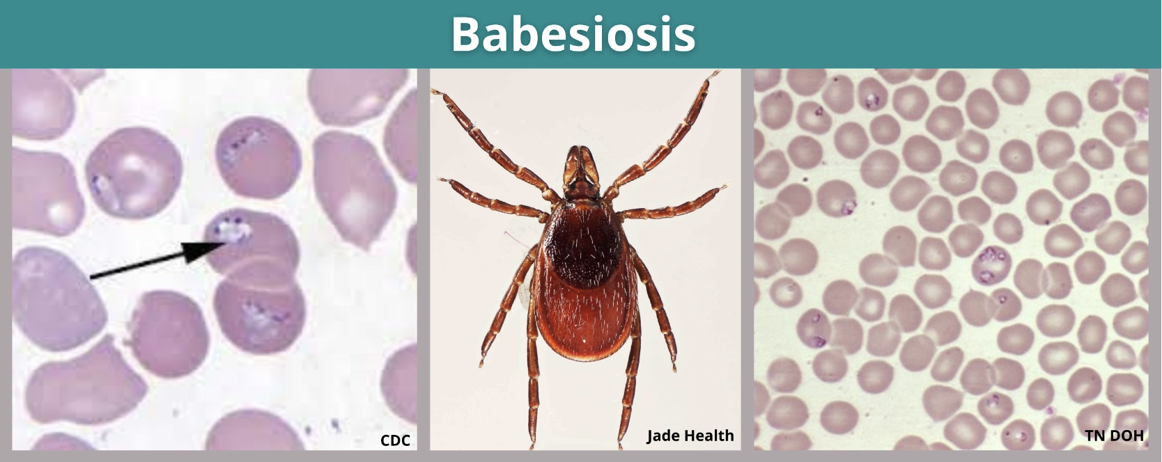
The most common symptoms include fever, muscle and joint pain, and headache. In certain patients, severe complications can occur, including thrombocytopenia, renal failure, and acute respiratory distress syndrome.
Thrombocytopenia is a condition characterized by abnormally low blood platelet counts. “Babesiosis can cause illness ranging from asymptomatic or mild to severe; the disease can be fatal, particularly among persons who are immunocompromised or asplenic.” Life Cycle of Babesia Microti (CDC)
The Babesia microti life cycle involves two hosts, which includes a rodent, primarily the white-footed mouse, Peromyscus leucopus, and a tick in the genus, Ixodes.
During a blood meal, a Babesia-infected tick introduces sporozoites into the mouse host. Sporozoites enter erythrocytes and undergo asexual reproduction (budding). In the blood, some parasites differentiate into male and female gametes although these cannot be distinguished at the light microscope level. The definitive host is the tick.
Once ingested by an appropriate tick , gametes unite and undergo a sporogonic cycle resulting in sporozoites. Transovarial transmission (also known as vertical, or hereditary, transmission) has been documented for “large” Babesia spp. but not for the “small” babesiae, such as B. Microti.
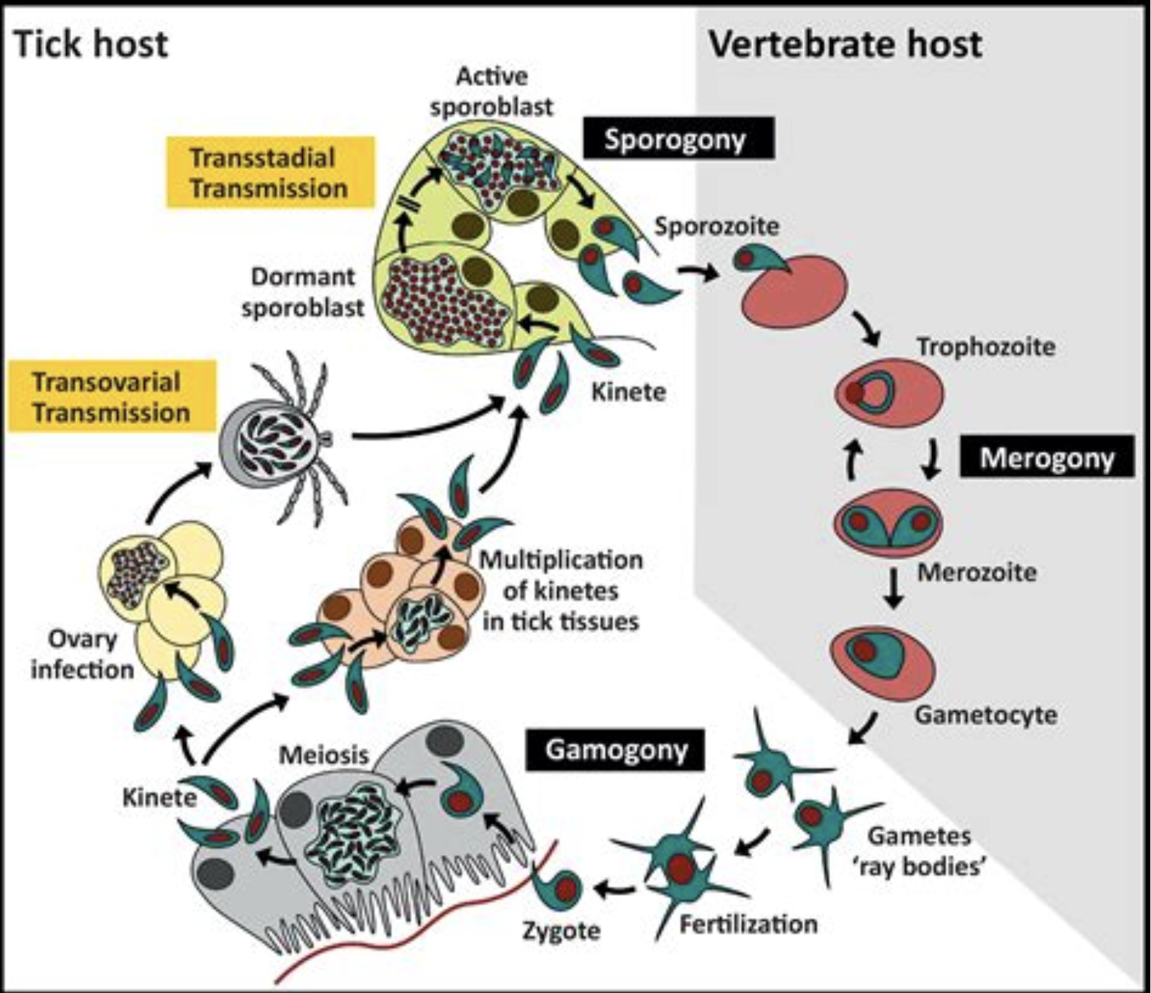
Humans enter the cycle when bitten by infected ticks. During a blood meal, a Babesia-infected tick introduces sporozoites into the human host. Sporozoites enter erythrocytes and undergo asexual replication (budding).
Multiplication of the blood stage parasites is responsible for the clinical manifestations of the disease. Humans are, for all practical purposes, dead-end hosts and there is probably little, if any, subsequent transmission that occurs from ticks feeding on infected persons. However, human to human transmission is well recognized to occur through blood transfusion.
Incidence
Between 2020 and 2024, on Marthas Vineyard 113 cases of Babesiosis were reported — a rate 11.6 times higher than the Massachusetts average. In Massachusetts the number of cases of human babesiosis trended upward from around 500 in 2016 to over 900 in 2025.
Laboratory Findings
Lab findings in Babesiosis typically include hemolytic anemia, thrombocytopenia, and elevated liver enzymes. A peripheral blood smear may reveal intraerythrocytic ring forms and tetrad formations known as Maltese crosses, characteristic of the infection.
FYI: The Maltese Cross was popular during the Christian crusades of the Middle Ages and has been adopted by modern fire fighters as a symbol of their profession.
Anemia results from decreased red blood cell count due to destruction of red blood cells. Thrombocytopenia can complicate the clinical picture. Increased levels of liver enzymes indicating liver involvement. While useful for epidemiological studies, serology alone is not reliable for diagnosing acute infections due to persistent antibodies. Polymerase Chain Reaction (PCR) can detect Babesia DNA and is particularly useful in cases of low parasitemia.
Pathophysiology
Alterations in RBC membranes cause decreased conformability and increased RBC adherence, which can lead to development of noncardiogenic pulmonary edema and acute respiratory distress syndrome (ARDS) among those severely affected. Fever, hemolytic anemia, and hemoglobinuria may result from Babesia infection. As with malaria, RBC fragments may cause capillary blockage or microvascular stasis, which could explain liver, splenic, renal, and central nervous system (CNS) involvement.
Animal studies have shown that increased cytoadherence of infected RBCs could cause these vascular blockages. As in malaria, cells of the reticuloendothelial system in the spleen remove damaged RBC fragments from the circulation. RBC destruction results in hemolytic anemia. The amount of hemolysis does not seem to be directly related to the degree of parasitemia, though the cause is unclear.
The spleen offers a critical host defense against babesiosis, as suggested by the higher incidence and greater severity of babesiosis in asplenic patients. The spleen traps the infected erythrocytes, and their ingestion by macrophages follows. Additionally, hypersplenism can lead to platelet sequestration which causes thrombocytopenia.
Complement activation by Babesia may lead to the generation of tumor necrosis factor (TNF) and interleukin-1 (IL-1). Decreased complement levels, increased circulating C1q-binding activity, and decreased C4, C3, and CH50 levels are observed in patients with babesiosis. The generation of these primarily macrophage-produced mediators may explain many of the clinical features, including fever, anorexia, arthralgias, myalgias, and the fulminant shock syndrome.
Treatment
Atovaquone works by inhibiting the mitochondrial electron transport chain in Babesia parasites, which is essential for their energy production and survival. Parasite Clearance: By disrupting their energy production, atovaquone helps clear Babesia parasites from the bloodstream during treatment. Azithromycin works by binding to the bacterial ribosome, disrupting protein synthesis
Treatment Regimen: Atovaquone is typically used in combination with Azithromycin for a standard treatment course of 7 to 10 days in immunocompetent patients. The first published documentation of successful treatment of this regimen appeared in the New England Journal of Medicine in November, 2000, by Dr. Peter Krause at Yale Medical School.